Common Indications for Bronchoscopy
- Unexplained Pneumonia :
Used to collect samples such as bronchoalveolar lavage (BAL), brushings, or biopsies from the affected lung areas seen on imaging.
- Suspected Tuberculosis :
Helpful in patients with dry cough or inconclusive sputum results. BAL and biopsies can help detect drug-resistant TB.
- Lung Cancer:
Used to collect samples from abnormal segments of the lungs as seen on CT/PET scans. Enlarged lymph nodes can be examined using TBNA (Transbronchial Needle Aspiration) or EBUS (Endobronchial Ultrasound).
Before the Procedure
- Inform your doctor about all medicines, supplements, or allergies.
-
You may be asked to stop taking blood thinners a few days before the test.
-
Avoid eating or drinking for a few hours prior to the procedure.
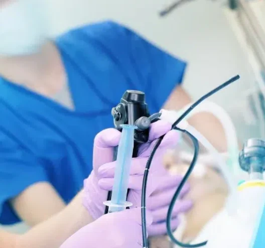
During the Procedure
- Usually done on an outpatient basis.
-
You'll lie on your back, and your nose, mouth, and throat will be numbed with a spray.
-
A mild sedative may be given through an IV. In some cases, general anesthesia is used.
-
If awake, the scope may trigger a temporary cough until the numbing takes full effect.
-
The procedure usually takes about 30 minutes, depending on what’s being done.
After the Procedure
- You'll be monitored for complications.
-
If sedated, you may not recall the test.
-
Eating or drinking is not allowed until the throat numbness wears off.
-
A sore throat or cough may persist for a day or two.
-
If biopsies were taken, results are usually available in a few days.
Uses of Bronchoscopy
Evaluation
Bronchoscopy can help diagnose:
- Persistent cough lasting over 3 months
-
Coughing up blood (hemoptysis)
-
Shortness of breath or unexplained low oxygen levels
-
Suspected airway obstruction
-
Tumors, lung scarring, or lung collapse seen on imaging
-
Lung infections not identified by other tests
-
Transplant rejection
-
Exposure to toxic gases or chemicals
EBUS (Endobronchial Ultrasound) may be used to locate and biopsy tumors deep in the lungs.
Newer diagnostic tools include:
- Autofluorescence bronchoscopy
-
Narrowband imaging
-
High-definition video bronchoscopy
Therapeutic Applications
Bronchoscopy can also be used to:
- Remove mucus or fluid
-
Extract foreign objects
-
Enlarge narrowed airways
-
Rinse out the bronchial passages
For some lung cancers near major airways, bronchoscopy can assist with internal radiation treatment (brachytherapy).
Types of Bronchoscopy
- Flexible Bronchoscopy
Most common type
Uses local anesthesia and mild sedation
- Rigid Bronchoscopy
Less common, used for more complex procedures
Requires general anesthesia and is done in an operating room